 Radiological examination are crucial for dental diagnostics. They allow to acquire inaccessible information on the state of dentition.
– Periapical X-rays
Periapical X-rays give detailed information about the state of a single tooth. They are known for its precision and the ability to reproduce even small details.
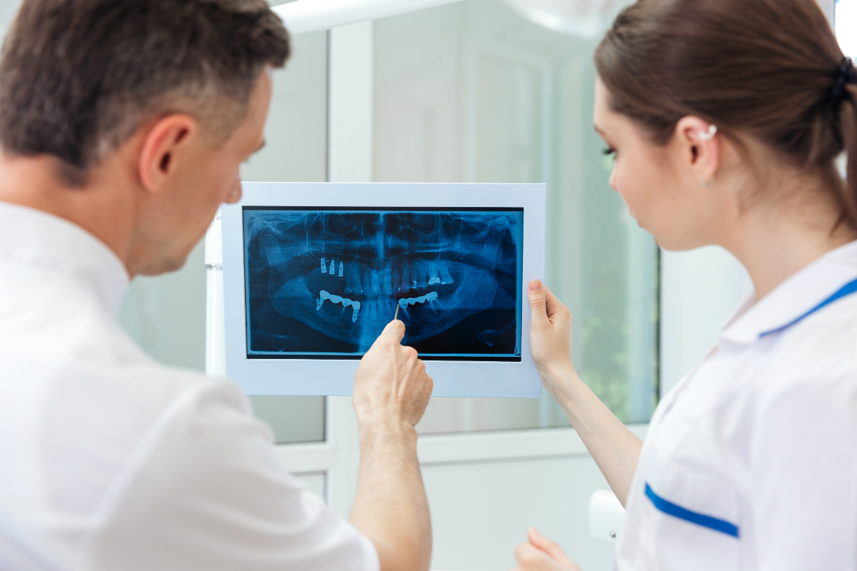
– Pantomographic X-rays
X-rays done in this method can show dentition, jaw bones and neighboring structures on one photo. They are less detailed than periapical X-rays, but allow doctors for comprehensive assessment of dentition. Currently pantomographic X-rays are used routinely before starting a dental treatment.
– Cefalometric X-ray
Cefalometric X-rays is used in, e.g. orthodontics. Such X-rays perfectly visualize facial skeleton’s profile. This allows to perform a very detailed diagnostics of occlusal abnormalities and design an accurate plan for orthodontic treatment.
-X-rays of temporomandibular joints
These types of X-rays are used in surgical dentistry, prosthodontics and orthodontics because all of those branches deal, to some extent, with treating diseases and temporomandibular joints abnormalities. Such examination brings important information on the cause, localization and types of disease symptoms, which are often ambiguous.